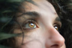
So we have two eyes side by side, giving us binocular stereoscopic vision. Each eye has a wide angle of view, and having them side by side gives us a wide angle of view. Each eye has a 150° field of view, and the two combined have a 180° field of view. The side-by-side arrangement of the eyes comes to us from evolution, as most predators have their eyes side-by-side, enabling us to appreciate distances. To appreciate the distance of an object, we need two distinct angles of view. Prey, on the other hand, will prefer the widest possible field of vision, even if this means limiting the binocular field of vision, i.e. in 3D, as some birds see 360°. It should also be noted that the two eyes operate independently.
In humans, each eye sees 90° outwards and 60° inwards. This gives a field of vision of 100° in 3D and 40° in 2D. The benefits of binocular vision are not limited to 3D vision, but that’s not all! Binocular vision also provides double information on the light emanating from a given point, and if this information is doubled, it enables us to perceive dimly-lit objects.
How does an eye work?
Well, like a camera’s darkroom. When light enters the eye, it passes through a first lens, the cornea, then a second lens, the crystalline lens. Between the two, light passes through the pupil, which lets in more or less light, like the diaphragm on a camera.
How does our pupil work?
In the dark, the pupil dilates to let in more light, a phenomenon we call mydriasis, and conversely the pupil contracts in the presence of light, a phenomenon we call miosis. Normally, this is a reflex, but sometimes it doesn’t go as planned. A case in point is David Johns, who was punched in the eye during a fight. This had the effect of keeping his pupil permanently open. The effect is called permanent mydriasis, and David Johns is better known as David Bowie.
Light rays that have passed through the cornea, the center of the pupil and the crystalline lens end up passing through the vitreous body – which makes up most of our transparent eye – and end up on the retina.
What is the retina and how does it work?
On the surface of the retina we find a host of sensors, cones and rods. Rods are specialized in acquiring information about light intensity, while cones are specialized in acquiring information about light frequency – cones are specialized in acquiring information about color. They are very bad at low light intensity, which is why we always see in black and white at night. Rods, on the other hand, are good at light intensity, even at low light levels, and bad at color perception, which is not their role.
The science of optics
In optics, we call night vision scotopic vision, from the ancient Greek “scotos” meaning darkness and “opsis” meaning sight. In fact, fear of the dark is called scotophobia.
Daytime or diurnal vision, photopic vision, from “photos”, light, and “opsis”, sight. At dusk or dawn we call the mesopic domain, from “meso” meaning the middle and “opsis”, sight.
Ocular performance?
Ocular performance is the performance of sight, but only at the level of the eye, without regard to the processing that the brain can make of it: the performance of the eye, considered as an instrument that captures light or images. There are 4 levels of information. There’s sharpness, resolution, contrast and movement.
- Sharpness: when we look at a point, we need as few points as possible in our eye. When we look at an unfocused dot, we have a multitude of information on the retina, enabling us to appreciate the larger dot. So a small dot needs to appear on the retina as small as possible.
- Resolution: the smallest distance we can distinguish in the image formed in the eye. In a camera or television, this is called a pixel.
- Contrast : this is the degree to which the image formed in the eye is able to distinguish the luminous intensity of two distinct surfaces in reality.
- Motion: how much the image formed in the eye will update when what we’re looking at is moving.
The cornea
For sharpness, what counts is the eye’s ability to focus light rays. In the human eye, the cornea is the first lens through which light rays pass. It’s a converging lens, so it’s thicker at the periphery and thinner at the center. It is only 1mm at the periphery and 0.5mm at the center. The cornea is responsible for 70% of the focusing of light rays. The cornea is normally transparent, but it can lose its transparency. To remain transparent, the cornea needs to bathe, remaining in a hydrated environment with 80% water per milligram of tissue. This is a vestige of our aquatic life, when we lived underwater.
The cornea is made up of 6 layers (the sixth layer was discovered in 2013). From the outer to the inner layer:
- The epithelium : 5 to 6 layers of cells thick, its main purpose is to properly distribute tears on the surface and protect the inside of the eye. This layer is in direct contact with the air and its impurities. This is also why the epithelium contains a mind-boggling number of nerve endings, whose sensitivity is purely painful. These are not sensors capable of giving information about heat, but only about pain. This explains why we can’t stand the slightest dust in our eyes. A surprising fact about the epithelium: its cells are completely renewed from the top layer to the base layer (basal layer). That’s why we can easily fit contact lenses without the slightest concern for the eye’s integrity.
- Bowman’s membrane: a membrane composed of collagen sandwiched between the epithelium and stroma. Its structure is totally random and blends into the stroma, making it difficult to distinguish. Its role is still very difficult to define. This membrane doesn’t renew itself, and damage to this layer leads to the appearance of scar tissue, which can cause visual disturbances.
- The stroma: the thickest layer of the cornea, representing 80% of its total thickness. It is made up of perfectly oriented, periodically striated collagen lamellae. It is mainly these lamellae that are responsible for the cornea’s good transparency. The stroma is also made up of keratocytes, the supporting cells responsible for renewing the collagen matrix, and a fundamental substance that ensures the collagen’s proper organization.
- The Dua layer : a difficult layer to discover, since it’s only a single cell in size: 0.015 millimetres. Its usefulness is uncertain. Its existence was only discovered in 2013.
- Descemet’s membrane: between 5 and 7 micrometers thick, it thickens over time. It is transparent and mainly composed of collagen.
- The endothelium : as with the epithelium, its main role is to act as a barrier inside the eye. It also controls stromal hydration. Its cells, with their honeycomb structure, literally pave the interior of the cornea.
Where do the light rays that have passed through the cornea go?
Light rays pass through the anterior chamber of the eye, the area between the cornea and the iris. The iris is the colored part surrounding the pupil. This anterior chamber is made up of aqueous humor, which is 90% water and other components such as chlorine and sodium, i.e. salt. The role of this liquid is to maintain a constant pressure in the eye in general, but also to nourish and repair the cornea. The pressure at this level of the eye increases over the years, and when this pressure becomes too great, glaucoma develops, progressively damaging the visual cells and causing progressive blindness.
On the other hand, light rays pass through the posterior chamber ofthe eye, the area between the iris and the lens. This, too, is made up of the aqueous humor secreted by the ciliary muscle, which enables the lens to move.
Le vitré
After the lens, we find the vitreous. A sort of gel, identical in composition to the cornea, it is composed of 90% water, collagen fibers and protein. Vitreous humor is the component of the vitreous and makes up 90% of the entire eye. Its role is to maintain the shape of the eye and press the retina against the back of the eye. Retinal detachment occurs when the aqueous humor slips between the retina and the pigmented epithelium, detaching the retina. If this membrane detaches, vision loss is immediate and may be irreversible. Surgical intervention within hours of the detachment’s appearance can save part of the visual field.
The lens
It is important for sharpness. This lens is capable of accommodation. Accommodation is the eye’s ability to see objects both near and far. With a diameter of 1 centimeter and half its thickness, the lens is held in place by the ciliary muscle, which, by contracting and dilating, modifies its shape to make it more or less curved. As a result, light rays passing through it focus on a more or less distant point.
What happens when light rays reach the eye?
Light generally travels in a straight line as long as it remains in the same medium. So light in air travels in a straight line, but as soon as it passes through another medium, a refraction process takes place: the ray is deflected. As it passes through the eye, light rays pass through several media and are refracted several times, ending up on the retina, projecting an image of what we’re observing outside. This is the principle of the darkroom.
When these rays converge on the retina, they activate the cone and rod photoreceptors, which send information about light intensity and frequency to the brain. Color and light intensity. To understand the notion of contrast and resolution, to have an image that is well defined and correctly marks light variations. We need to look at how the cones and rods are distributed on the surface of the retina.
Directly in front of the pupil, at the back of the eye, lies a cone-dense zone called the macula, 5mm in diameter. This is precisely the most cone-dense area of our eye, and within it lies the even more cone-rich fovea. Because this zone is so cone-rich, it is able to transmit a great deal of color information. This zone has a high resolution, but beware, as this zone has a high concentration of cones and not rods, we have a high resolution in the photopic period, during the day. In the dark, this zone doesn’t work very well… In the fovea, there are no rods and its structure is quite astonishing, a honeycomb pattern of cones. It has been mathematically proven that honeycomb paving is the optimum paving for covering the largest surface area with the smallest joint perimeter.
As we move away from the macula, we find more and more rods and less and less cones. This means that, in terms of ocular performance, right at the center of the pupil, at the center of our vision, we have an area that is really high-resolution and high-contrast. As we move away from the center of our vision, we lose resolution and contrast. Note that we won’t lose sharpness. On the contrary, we’ll have better information on light intensity. That’s why we can easily make out the flashing lights of police cars in our field of vision.
How do we detect movement with our eyes?
When the resolution is fine enough to detect reasonable movement (under the right conditions), the question of movement is purely a question of image refreshment. How fast is the eye able to refresh the image and send it to the brain? For a camera, this is called FPS, but for the eye it’s a little more different: it’s how often the cone or rod is able to acquire new information and transmit it to the brain. We’re capable of watching films of 25 to 30 frames per second on TV or YouTube. What we observe is that a subject confronted with a stroboscopic light of 50 to 60 flashes per second sees a persistent light.
What is the blind spot or papilla of our eye?
In the retina, there is an area of the eye where the optic nerve is inserted and where blood vessels arrive and leave. Mechanically, there can be no photoreceptors in this area.
Eyes and ultraviolet rays
On the retina we have cone and rod photoreceptors. The rods contain a protein pigment, Rhodopsin , which is sensitive to ultraviolet light. If our crystalline lens combined with the vitreous did not absorb ultraviolet light, we would be fully able to see it. This is a frequency that rods shouldn’t normally see, as they don’t perceive colors…


